Bone maintenance with Dentalis System™
Bone maintenance with Dentalis System™
Bone maintenance with Dentalis System™
Time to challenge old facts
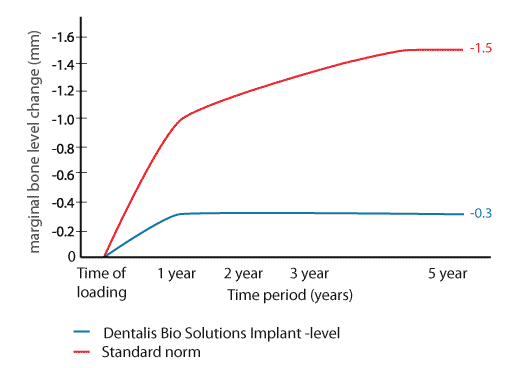
How do you develop the best long-term treatment results for your patients? The norm for dental implant treatment success from the 1990′s does not reflect what we can achieve today. There is no reason why the patient or clinician should accept a bone loss of up to 1.5 millimeters based on a standard set 25 years ago around the neck of the implants.
As a company that understands the importance of research and development, Dentalis Bio Solutions has monitored the maintenance of the cortical bone level around our own systems’ for many years, and noted that the cortical bone levels around Dentalis implants have been very well kept.
When comparing our exceptional result to the current standard norm, it turns out that there is a huge gap between what can be achieved with our system and what is currently accepted as a successful treatment result.
.
Dentalis Implant System™ level
Dentalis Bio Solutions conducted a review of the literature analyzing the Dentalis Implant System and resulting changes in bone level. A total of sixteen prospective, full cohort studies reporting on implant treatment and applying a standard protocol were reviewed, and the conclusion is that the Dentalis Implant System has an average marginal bone level reduction of less than 0.4 mm after the first year of loading and remains stable after five years – a result that is five times better compared to the current standard norm.
For over two decades, Dentalis has led implant dentistry into a new era of attainability and aesthetics.
The pink tissue versatile implant neck combines superior
gingival aesthetics and high primary stability,
improved placement, temporization,
which is particularly beneficial in the
most aesthetically demanding cases.
REFERENCES:
1.Bittner N, Schulze-Späte U, Cleber S, Da Silva J, Kim D, Tarnow D, Ishikawa-Nagai S, Gil M. Comparison of Peri-implant Soft Tissue Color with the Use of Pink-Neck vs Gray Implants and Abutments Based on Soft Tissue Thickness:
A 6-Month Follow-up Study. Int J Prosthodont. 2020Jan/Feb;33(1):29-38.
2. Gil M, Ishikawa-Nagai S, Elani H, Da Silva J, Kim D, Tarnow D, Schulze-Späte U, Cleber S, Bittner N. Comparison of the Color Appearance of Peri-implant Soft Tissue with Natural Gingiva Using Anodized Pink-Neck Implants and Pink Abutments: A Prospective Clinical Trial. Int J Oral Maxillofac Implants. 2019 May/June;34(3):752–758.
3.Gil M, Ishikawa-Nagai S, Elani H. A prospective clinical trial to assess the optical efficacy of pink neck implants and pink abutments on soft tissue aesthetics. J Esthet Restor Dent. 2017;29(6):1-7.38.
For over two decades, Dentalis has led implant dentistry into a new era of attainability and aesthetics.
The pink tissue versatile implant neck combines superior gingival aesthetics and high primary stability, improved placement, and temporization, which is particularly beneficial in aesthetically demanding cases.

REFERENCES:
1.Bittner N, Schulze-Späte U, Cleber S, Da Silva J, Kim D, Tarnow D, Ishikawa-Nagai S, Gil M. Comparison of Peri-implant Soft Tissue Color with the Use of Pink-Neck vs Gray Implants and Abutments Based on Soft Tissue Thickness:
A 6-Month Follow-up Study. Int J Prosthodont. 2020Jan/Feb;33(1):29-38.
2. Gil M, Ishikawa-Nagai S, Elani H, Da Silva J, Kim D, Tarnow D, Schulze-Späte U, Cleber S, Bittner N. Comparison of the Color Appearance of Peri-implant Soft Tissue with Natural Gingiva Using Anodized Pink-Neck Implants and Pink Abutments: A Prospective Clinical Trial. Int J Oral Maxillofac Implants. 2019 May/June;34(3):752–758.
3.Gil M, Ishikawa-Nagai S, Elani H. A prospective clinical trial to assess the optical efficacy of pink neck implants and pink abutments on soft tissue aesthetics. J Esthet Restor Dent. 2017;29(6):1-7.38.
Standard norm
The red line in the graph illustrates what has, until now, been perceived as the regular norm for acceptable bone loss related to dental implant treatment. The figures are based on the three different articles stated next to the graph above. The articles describe the acceptable bone loss to be less than 1.0 mm during the first year of loading and less than 0.2 mm annually during the following year. Thereafter, the marginal bone loss tapers off, resulting in a marginal bone loss of approximately 1.5 millimetres after five years of loading.
*Regular protocol means treatment without immediate loading or/and without bone augmentation.